
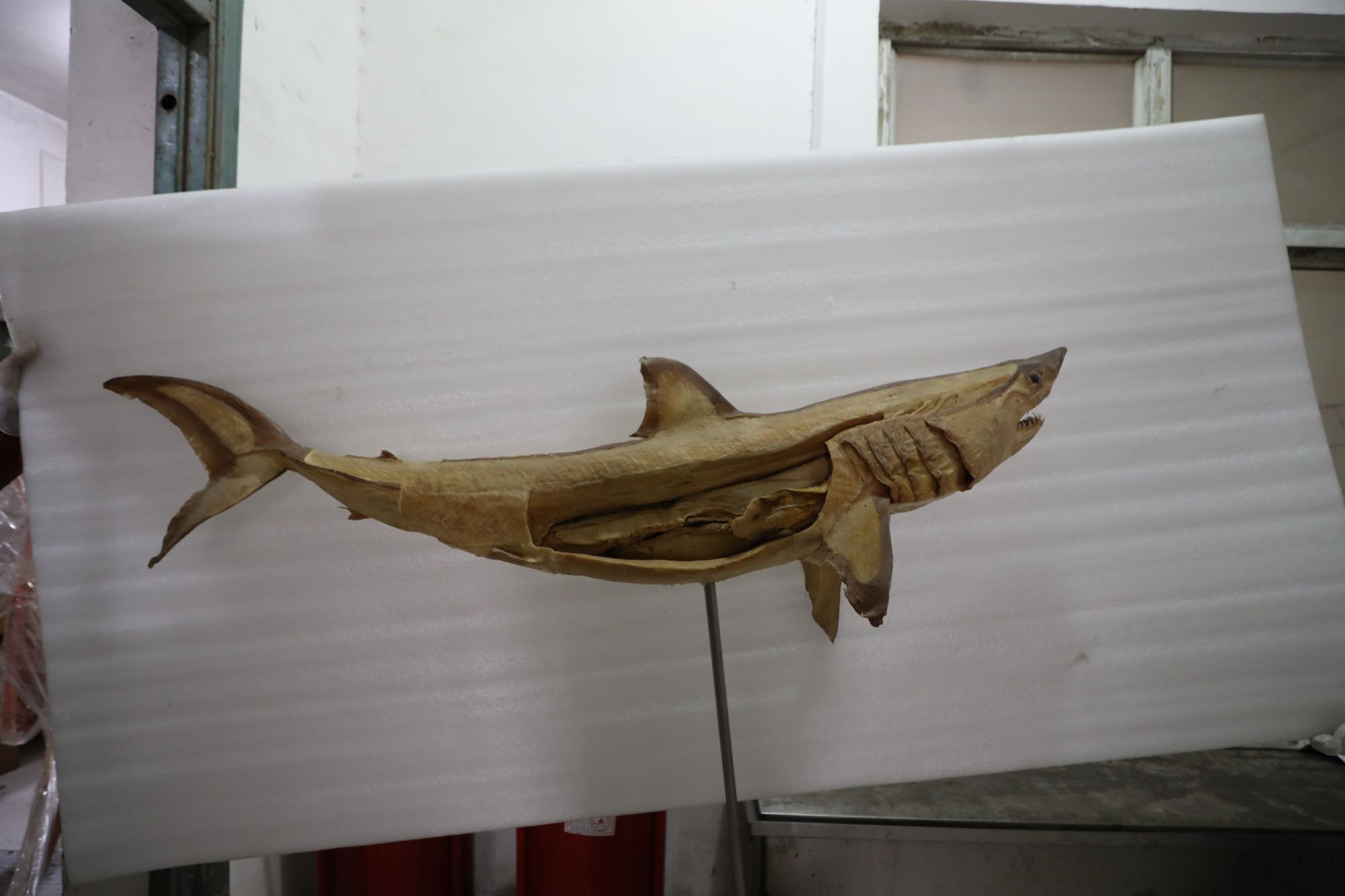
Plastinated specimens play a crucial role in education, especially in animal life science museums, breaking the limitations of traditional teaching models. By showcasing the intricate structures of animal internal organs, muscles, and bones, plastinated specimens provide learners with an unprecedented intuitive experience. For example, in anatomy classes, students can clearly observe the respiratory system of a whale shark or the digestive organs of a giant panda. This "high-definition photograph" level of detail makes abstract biological knowledge tangible. This intuitiveness not only improves teaching efficiency but also stimulates students' interest in life sciences and cultivates their observation and reasoning abilities.
Furthermore, the long-term preservation properties of plastinated specimens make them a stable teaching resource. Compared to the defects of traditional specimens, such as deformation or fading, plastination ensures the durability of specimens, providing reliable support for repeated learning and research. This innovation has promoted the deepening of education from theory to practice, helping learners to understand animal physiology and ecology more deeply.
At the scientific research level, plastinated specimens are important tools for exploring animal morphology and function. Scientists use these specimens for anatomical studies, analyzing species' evolutionary adaptations, behavioral pattern

