
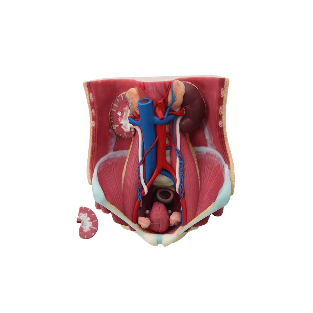
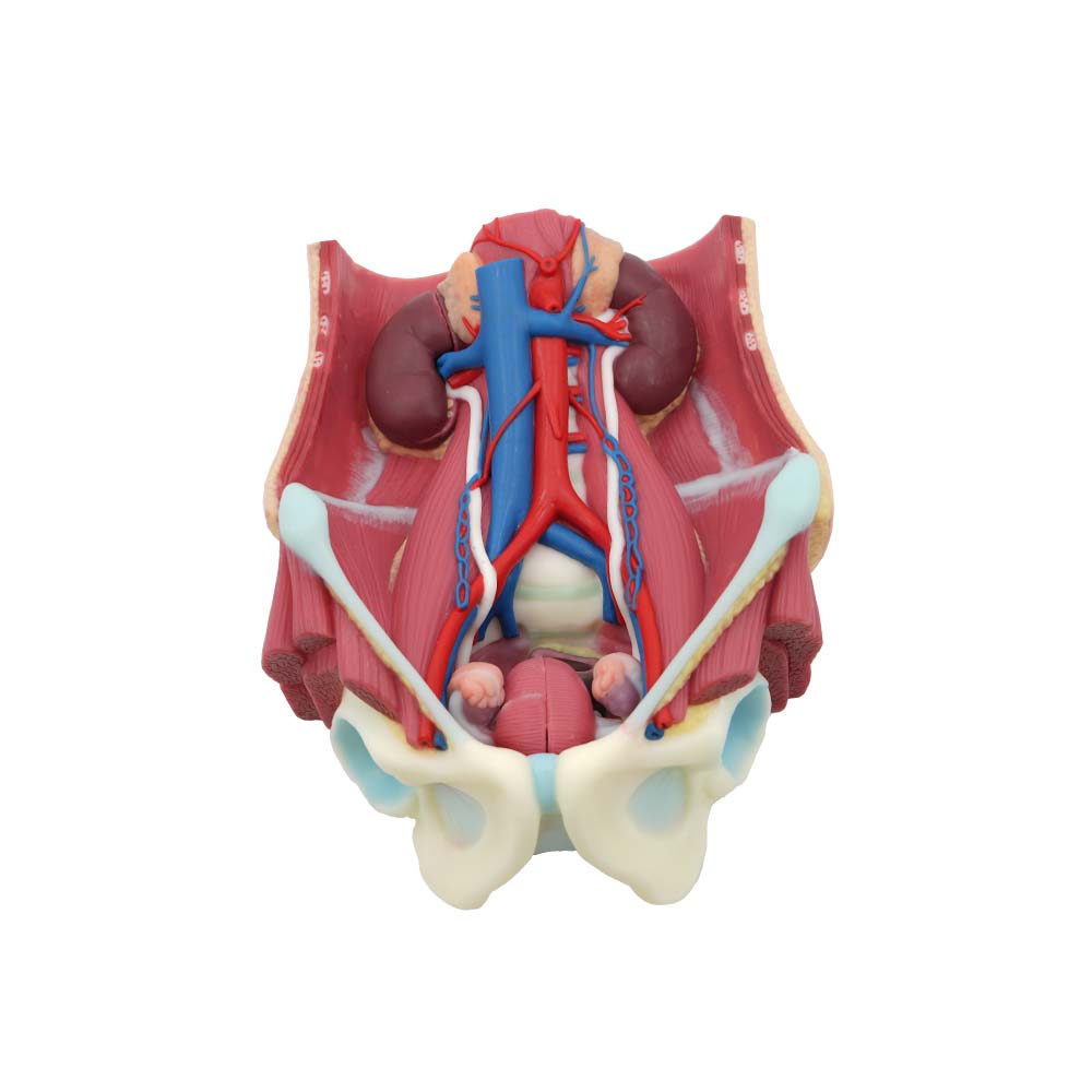
Based on real human anatomy, the models visually demonstrate the spatial location of organs (such as the retroperitoneal location of the kidneys and the pelvic proximity of the bladder) and their internal structures (such as the distribution of nephrons and the physiological narrowing of the ureters). This helps students develop three-dimensional spatial thinking, avoiding the cognitive barriers of "mental modeling" based on two-dimensional diagrams.
Meiwo human urinary system anatomy model primarily displays the various organs of the urinary system, the internal anatomical structure of the kidneys in cross-section, the internal and external anatomy of the male and female bladders, and the anatomy of the posterior abdominal wall muscles. The human urinary system anatomical model is made of environmentally friendly, food-grade soft silicone material, free of heavy metals such as lead, mercury, cadmium, hexavalent chromium, soluble antimony, soluble arsenic, soluble barium, soluble lead, soluble mercury, and soluble selenium, ensuring product stability and durability. It is non-toxic and odorless. The model boasts accurate structure and lifelike appearance. It can be repeatedly bent and cleaned, and is durable, drop-resistant, and easy to disassemble and assemble. The anatomical structures are created using a one-piece injection molding technique. Arteries (red), veins (blue), and nerves (yellow) are filled and molded in a single piece using color separation, ensuring no fading. Muscles and organs are also injection molded in a single piece according to their location (not computer-generated or painted). The anatomical structures are detailed, reaching 3-4 levels of branching.
The human urinary system anatomy model supports disassembly and reconstruction (such as a replaceable external genitalia module) to adapt to different learning progress. Instructors can provide customized feedback based on student performance data, reinforcing weak areas. Compared to real surgical demonstrations, the model is reusable and has low maintenance costs, while avoiding ethical controversies and patient privacy issues, making it a widely accessible teaching resource for medical schools.
By visually representing the precise logic of life sciences, the urinary system anatomical model not only overcomes the traditional teaching dilemma of "emphasizing theory over practice" but also serves as a bridge for cultivating clinical thinking and humanistic care. Its value in deepening anatomical understanding, honing operational skills, and integrating interdisciplinary knowledge will continue to drive medical education toward precision and humanization.

