
Traditional teaching methods using formalin-soaked foot specimens have inherent drawbacks, including a pungent odor, tissue atrophy and hardening, color fading, easy spoilage and damage, and inability to be reused long-term. Furthermore, physical specimens are often in fixed positions, preventing free movement and touch, and some learners may develop a psychological aversion to them due to their special nature, affecting teaching effectiveness. High-fidelity foot anatomy models, on the other hand, are odorless, wear-resistant, and have a long shelf life. They allow learners to observe closely, identify structures by touch, and flexibly adjust joint angles to simulate normal foot movement. They can be repeatedly used for classroom demonstrations and group training, adapting to routine teaching scenarios such as daily classroom instruction and laboratory training, effectively reducing the cost of teaching materials.
The models are suitable for various teaching methods, including classroom demonstrations, group teaching, and student self-directed hands-on practice. Learners can touch and identify various foot structures, adjust joint angles, and trace the course of nerves and blood vessels, facilitating in-class assessments, structural identification exercises, and simulations of movement mechanisms. This hands-on teaching model enhances learners' practical skills and cultivates precise structural identification abilities. It also focuses classroom attention, stimulates active observation and inquiry, effectively solidifies fundamental medical anatomy skills, and improves teaching and learning effectiveness.
High-fidelity foot anatomy models can serve as foundational teaching aids for pre-course courses in orthopedics, hand and foot surgery, rehabilitation medicine, and sports medicine. Through these models, the anatomical basis of common foot and ankle fractures, dislocations, ligament injuries, plantar fasciitis, flat feet, and high arches can be intuitively explained. Learners can understand the connection between diseases and foot anatomy, and comprehend the logic behind clinical diagnosis, treatment, and rehabilitation plans. Simultaneously, the models can simulate basic clinical skills such as clinical examinations and reduction procedures, laying a solid anatomical foundation for subsequent clinical internships and procedures. This facilitates a smooth transition from basic anatomical knowledge to clinical application, aligning with the entire medical student training system.
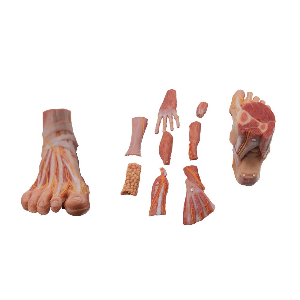
High-fidelity foot anatomy models are replicas of real human feet, featuring detailed structures and lifelike textures. Their core role in medical education is to address the shortcomings of traditional teaching methods and improve teaching quality: First, they transform abstract foot anatomy into three-dimensional, intuitive physical objects, helping learners quickly develop a three-dimensional spatial concept and overcome the learning difficulties associated with confusing bone, joint, and ligament structures. Second, they accurately reproduce all the fine structures of the foot, meeting the needs of refined teaching and practical training, and overcoming the drawbacks of formalin specimens such as odor, perishability, and immobility. Third, they enable a coherent explanation of anatomical structures, physiological functions, and movement mechanisms, moving beyond rote memorization and deepening understanding. Fourth, they enhance hands-on interaction, adapting to classroom demonstrations and group training, solidifying fundamental anatomical skills and structural identification abilities. Fifth, they connect with clinical courses, bridging basic anatomy with foot and ankle clinical practice and rehabilitation medicine, while also being suitable for multi-scenario teaching and popular science, expanding their application value.
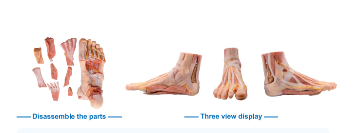
This highly realistic foot anatomy model meticulously replicates the morphology and structure of a real human foot, accurately reproducing all core details such as bones, joints, ligaments, and nerves and blood vessels, and intuitively presenting the spatial structure and movement mechanism of the foot. In teaching, it clearly demonstrates the physiological functions and anatomical connections of the foot, overcoming the inherent limitations of traditional physical specimens. It achieves integrated teaching of anatomical structure, physiological function, and clinical foundations, adaptable to various scenarios such as classroom demonstrations, group training, pre-clinical teaching, and popular science presentations. It reduces learning difficulty and improves memory efficiency, while also contributing to teaching quality and practical skills enhancement, making it a core training tool for basic medical education and pre-clinical training.

