
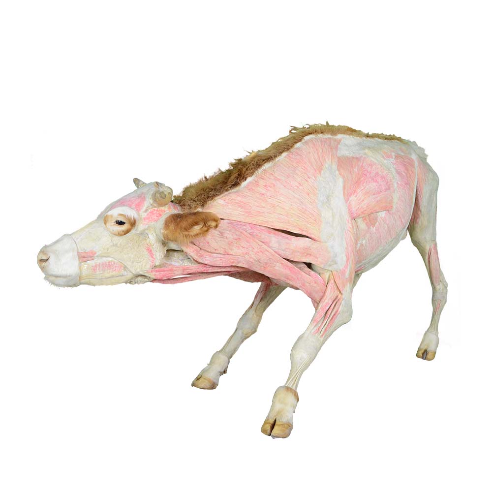
Cattle body plastination specimens are teaching aids made using advanced bioplasticization technology. Their core advantage lies in their ability to completely preserve the cattle's morphology, organ location, and tissue details from its pre-death state. They are also non-toxic, odorless, directly tactile, and durable, effectively compensating for the shortcomings of traditional teaching methods and playing an irreplaceable role in the teaching of veterinary medicine, animal science, and animal husbandry.
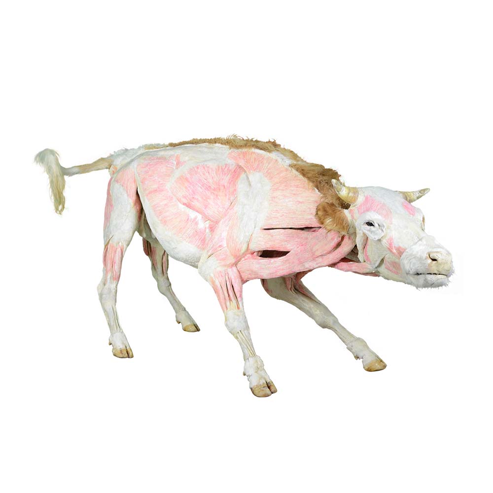
The core difficulty in courses such as animal anatomy and animal physiology lies in the abstract structure and physiological mechanisms of the organism. Traditional teaching often relies on textbook illustrations, two-dimensional videos, or fragmented specimens, making it difficult for students to develop three-dimensional spatial cognition, often resulting in rote memorization without the ability to apply knowledge flexibly. Cattle body plastination specimens can visually present the complete structure of the cattle body's systems, from the branching of blood vessels and nerves and the physiological texture of organs to the spatial relationships between systems and the connections between organs, all clearly discernible, making abstract theoretical knowledge concrete and perceptible.
For example, when explaining the cattle ruminant digestive system, students can directly observe the morphology, size, location, and connection of the rumen, reticulum, omasum, and abomasum to the esophagus and intestines using a complete plastinated specimen, gaining an intuitive understanding of the unique physiological structure of ruminants. When learning about the circulatory system, the clearly presented network of arteries and veins on the specimen allows students to quickly grasp the blood flow path and substance exchange mechanism, effectively overcoming the "seeing through a veil" learning dilemma and helping students build a systematic and complete knowledge framework, achieving a deep connection between theoretical knowledge and actual bodily structures.
The cattle body plastination specimen is highly versatile and can be adapted to multiple courses and various teaching scenarios, providing strong support for innovative teaching models. In classroom demonstrations, teachers can use the specimen to visually explain key and difficult points, allowing students to observe structural details from different angles through rotation and touch, enhancing classroom interaction. In group learning, students can discuss the specimen, exchange observations, raise questions, and collaboratively explore solutions, cultivating teamwork and self-learning abilities.
Furthermore, with the development of digital teaching, cattle body plastination specimens can be deeply integrated with modern technology to construct a three-dimensional teaching model of "physical specimen + digital resources." By scanning the QR code accompanying the specimen, students can access corresponding 3D anatomical models, knowledge point analyses, clinical case videos, and other digital content, achieving a linked learning approach of "physical observation + online extension." Teachers can use the specimens to conduct virtual simulation experiments, guiding students in virtual dissection operations, with the system providing real-time error correction and guidance, constructing a closed-loop teaching system of "theoretical learning - physical observation - virtual practice - assessment," improving teaching efficiency and relevance. Simultaneously, the specimens can also serve as popular science resources for youth study tours or public science education, helping to popularize knowledge about animal husbandry, animal health, and other related topics, highlighting the characteristics of the subject's teaching.
The lifelike appearance and realistic texture of cattle body plastination specimens effectively attract students' attention, stimulating their curiosity and desire to explore, changing the traditionally dull and tedious state of anatomy teaching. By touching and carefully observing the specimens, students actively explore the functional relationships between various systems and organs, cultivating their ability to analyze and solve problems through observation and reflection. For example, when observing specimens, students may proactively ask questions such as "the functional differences of muscles in different parts" and "the correlation between blood vessel distribution and disease diagnosis and treatment," actively participating in knowledge exploration and gradually developing rigorous scientific thinking and inquiry skills.
Whole-body plastinated bovine specimens, with their advantages of being realistic, intuitive, safe, durable, and highly adaptable, effectively compensate for the shortcomings of traditional teaching methods. They play an important role in bridging theory and practice, improving practical skills, innovating teaching models, supporting scientific research assessments, and stimulating learning interest. They are an important teaching tool for high-quality teaching and professional training in animal husbandry and veterinary medicine, and are of great significance for promoting the development of discipline teaching and contributing to the sustainable development of animal husbandry.

