
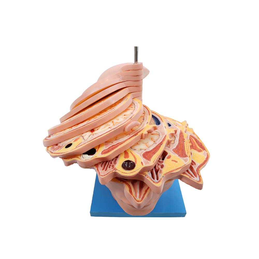
1. Visually Demonstrating Human Structure and Precisely Transmitting Knowledge
Human cross-sectional anatomy models can accurately demonstrate key and challenging aspects of anatomy. Through the model, one can see the structures in different planes, such as the transverse, sagittal, and coronal sections of the human body. This is like observing the human body sliced into pieces; students can visually see the location, shape, size, and adjacent relationships of organs with surrounding tissues at each cross-sectional level. For complex anatomical structures, such as the neurovascular foramina at the base of the skull and the adjacent relationships of organs in the pelvic cavity, the model can clearly present these structures
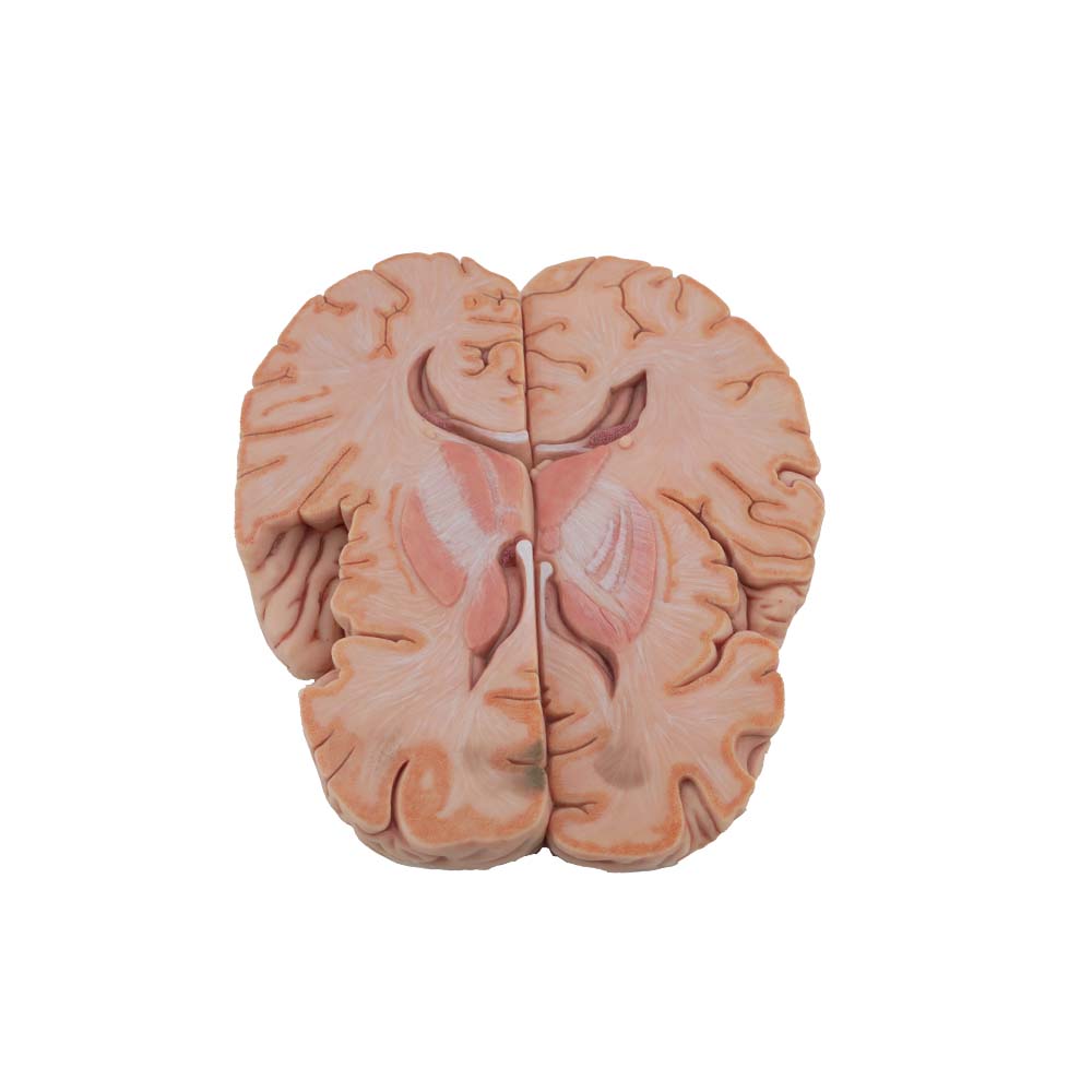
1. Teachers can directly point to specific parts of the model during explanation. For example, when explaining cerebral blood vessels, a brain sectional model allows teachers to accurately point out the pathways of blood vessels such as the middle cerebral artery and anterior cerebral artery within the brain parenchyma, as well as their relationship with surrounding brain tissue. This intuitive teaching method enables students to understand and master these key knowledge points more quickly.
2. Aiding Understanding of Abstract Concepts and Rapid Knowledge Absorption
Compared to simple textual descriptions and two-dimensional images, anatomical models of the human body are more easily accepted and understood by students. Research shows that combining visual and tactile senses can significantly improve learning outcomes. While observing the model, students can touch and feel the texture and shape of different structures; this multi-sensory learning method accelerates knowledge absorption.
For example, when learning about hand anatomy, students can observe a hand sectional model to see the distribution of flexor tendons, extensor tendons, nerves, and blood vessels at different finger levels. Simultaneously, they can touch the model to feel the direction of tendons and the thickness of nerves, thus gaining a deeper understanding of the intricate anatomical structures of the hand and mastering more knowledge in a shorter time.
Meiwo Science's anatomical cross-sectional models integrate virtual reality technology and are equipped with QR code labels, allowing users to scan the QR codes with their mobile phones to view similar physical models or specimens.
Students can scan the model's QR code with their mobile phones to access similar physical models or specimens anytime, anywhere, embarking on an immersive learning journey. The mobile app allows for 360-degree rotation, flipping, panning, and free zooming and dragging, completely breaking the constraints of traditional two-dimensional teaching and allowing students to examine complex anatomical details from any angle. Automatic annotation (in both Chinese and English) and voice broadcasting work together, making it easy to memorize obscure professional terminology and recognize precise anatomical locations. Lock and reset functions help students focus on key areas and study them repeatedly, while the drawing function meets students' needs for personalized marking and note-taking. Combined with infinitely changeable background colors, it creates a comfortable and focused learning atmosphere. Particularly thoughtful is the optimal viewing angle function; by clicking on the name in the annotated structure directory, students can instantly and accurately locate the corresponding structure to the best field of view, greatly improving learning efficiency. Meanwhile, the annotation and assessment function for structural identification and recognition practice questions provides students with a "practice ground," allowing them to manually annotate and compare their work with the correct answers, enabling real-time testing of learning outcomes and reinforcing knowledge mastery.
Human sectional anatomy models can simulate scenarios from actual clinical operations. In surgical teaching, models can demonstrate the anatomical layers through which a surgical incision passes. For example, in an abdominal surgery model, students can see the complete anatomical layers from the skin, subcutaneous tissue, fascia to abdominal organs, understanding the blood vessels, nerves, and other important structures they may encounter during surgery.
This helps cultivate students' clinical operational awareness, giving them a certain intuitive understanding of surgical procedures before entering clinical practice, familiarizing them with surgical pathways in advance, and reducing errors in actual operations.
For disease diagnosis, understanding human sectional anatomy is crucial. In imaging teaching, comparing human sectional anatomy models with CT, MRI, and other imaging data helps students better interpret images.
For example, when learning about lung disease diagnosis, students can compare chest sectional anatomical models with lung CT images to understand the differences in sectional structure between normal and diseased lung tissue, such as the morphology and location of lesions like pulmonary nodules and inflammation at different levels. This improves their diagnostic abilities and lays a solid foundation for future clinical work.
Human sectional anatomical models, as an advanced teaching tool, have become an indispensable part of medical education. They not only enhance students' mastery of anatomical knowledge and clinical skills but also promote technological innovation and improve the quality and effectiveness of medical education. With continuous technological advancements, the application prospects of sectional anatomical models will be even broader.
Meiwo Science focuses on the production and sales of medical anatomical models, primarily including human anatomical models, high-fidelity animal models, and PVC models. These products possess several important characteristics and advantages, including accurate anatomical landmarks, lifelike morphology, aesthetically pleasing appearance, durability, non-toxicity, odorlessness, impact resistance, and easy assembly/disassembly. Furthermore, Meiwo Medical Educational Models are equipped with digital functionality, allowing users to scan QR codes for online viewing and learning. Hongyu Medical Education's medical models not only meet the diverse needs of medical education and clinical practice, but these models can also help medical students better understand human structure and function, improve clinical skills, and promote advances in medical research.

